Structure and function of the spine
The evolutionary feature of a person is upright posture. For this advantage, it is necessary to pay, since the load on the spine is shifted by about ninety degrees and turns out to be directed from the top down. In this case, the spine acts as the supporting body of the trunk. Mobility of the trunk should be somewhat limited, in order to avoid the underside of excessive flexibility. In addition to this dilemma, a compromise between stability and mobility, the spine has an important function as a bone protective shell for the spinal cord.
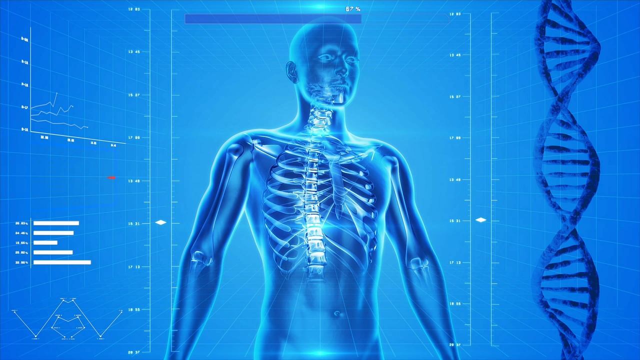
The spine itself, when viewed from the side, has a double S-like bend and is divided into the cervical, thoracic and lumbar divisions.
The sacrum (sacral bone), which consists of fused vertebrae, is adjacent to the lumbar spine. Elements of the spine are single vertebrae, which usually a person has 24 (seven cervical, twelve thoracic and five lumbar vertebrae). The uppermost vertebra is connected by several joints to the base of the skull. To the thoracic vertebrae, ribs are attached. The lower lumbar vertebrae borders on the sacrum, which in turn is connected to the pelvis.
The vertebrae are not simply stacked on each other, but are interconnected in the anterior parts by cartilaginous plates – the intervertebral discs – and in the posterior sections to the left and to the right – with the help of joints. The ligaments and the muscular frame complete the joints and allow the spine to perform only certain movements. Between the two vertebrae to the right and left come spinal nerves and with its branches reaches the periphery, from the cervical and lumbar spine to the fingers and toes (see image). The intervertebral discs themselves undergo a premature aging process and their nutrition depends on the shift between load and relaxation. They can perform their buffer function when loaded most fully when the load is evenly distributed along the body. The forces should be directed mainly from the head to the legs, and the asymmetric load, for example, when lifting the load with the bent back, should be avoided. It also has a negative impact on the metabolism of the intervertebral disk, unilateral loads, for example, in the form of prolonged sitting or activities in so-called forced postures. It is necessary to give preference to changes in body postures.
Diseases of the intervertebral discs – national sickness number 1
In medical practice, intervertebral disk diseases are the most common single diagnosis. Almost every person in industrialized countries faces throughout this life with this. In almost 2/3 cases of disease, lumbar vertebrae are affected.
Cervical vertebrae are affected approximately in 1/3 of cases, diseases are rare. Most often, the disease of the lumbar vertebra begins at the age of between 30 and 60 years, cervical vertebrae – a little later.
Causes of pain
Nutrition of intervertebral discs is regarded as an extremely important process, since discs do not have their own feeding blood vessels, and nutrients are forced to be obtained from surrounding tissues. With loads such as, for example, standing upright, sitting or lifting the load, the liquid is squeezed out of the intervertebral disc, releasing metabolic products. In addition, vice versa – when relaxing (especially when lying), the discs absorb liquid and nutrients from the surrounding tissues. Such limited exchange processes cause early wear processes, often detected by the age of thirty. Intervertebral discs weaken, cracking occurs. With asymmetric loads, the disc core can move, manifested by prolapse (bulging) or a herniated disc. Symptoms occur only with prolapses or herniated intervertebral discs directed back, by applying pressure on the ligamentous apparatus that has pain sensitivity, or on the nerves. Still, pain can occur and without dislocation of intervertebral discs. The strongly compressed disc is reduced in vertical size, and small intervertebral joints approach each other and squeeze together. In addition, aging of other joints of the body, called arthrosis, can occur. As a result, the spinal canal can be narrowed, the lack of space can lead to the appearance of symptoms; most often this occurs in older age.
Painful sensations often cause a muscle contraction, which in turn leads to new pain. Thus, some pains strengthen others, and vice versa, a vicious circle is formed. The picture of the disease can amplify itself.
As a natural reaction to wear occurs as hardening of the disc, also called “beneficial hardening”, due to the loss of water. Because of this, the risk of hernia formation gradually decreases.
Symptoms
Symptoms depend on which structures are damaged
The pain may have different localization.
Pain in diseases of the cervical spine:
Pain in the neck
Irradiation of pain in the back of the head
Irradiation of shoulder pain
Irradiation of pain in the hands/fingers
Pain in diseases of the thoracic spine:
Back pain
Irradiation of shingles along ribs
Pain in diseases of the lumbar spine:
Deep back pain
Irradiation of pain in the buttocks or thigh
Irradiation of pain in the shin or foot
In addition to pain, various functional impairments of the function of the spinal nerves can occur.
This can be manifested, for example, in the form of itching, numbness or weakness or muscle paralysis.
Massive herniated intervertebral discs with complete paralysis and disruption of the bladder or rectum are very rare.
Diagnostics
With the first symptoms, the attending physician conducts a thorough physical examination of the spine, which also includes a superficial check of the function of the nerves. Standard radiography serves to exclude rare diseases (displacement of vertebrae, inflammation, tumor diseases), which can cause similar symptoms. This method can detect the wear of the intervertebral disc, but not its hernia.
With neurologic functional disorders, as well as with persistent symptoms, a special layer-by-layer examination is performed.
Computed tomography (CT) makes it possible to obtain good-quality data on bone structures. Intervertebral disks and other soft tissue structures are better visualized by magnetic resonance imaging (MRI). Measurement of nerve conduction velocity and special laboratory tests (including rheumatic tests) may be necessary to clarify the diagnosis.
Treatment
The choice of the method of treatment depends on the course of the disease and the apparent symptoms. For acute pain, appropriate analgesics are prescribed in the tablet, injection or infusion forms. As soon as the patient's condition permits, he is prescribed exercise therapy. “School back” complete the activities to prevent relapses.
In case of chronic disorders, such measures should be carried out for a long time.
At the same time, the decisive factor here is strengthening the muscles of the back and abdomen, as well as changing the way of life. Do not take long-term pain medication.
Some patients with surgery for intervertebral disc disease require surgery. Such measures should be carried out firstly with acute signs of paralysis due to herniated discs, when the diagnosis is confirmed by MRI and qualified neurologic examination. In addition to the open operation on the intervertebral disk, it is also possible to perform the operation with the help of an operating microscope, there are also other methods that are to some extent minimally invasive, that is, the surrounding tissues are affected as little as possible.
With a small cut through the skin through a small hole between the arcs of the vertebrae, you can remove the herniated intervertebral disc.
Thus, the early-ligated spine of the spinal nerve is released.
Postoperative care in the treatment of a herniated disc
After an early postoperative phase and transfer back to the general department, conservative therapeutic exercise and patient remobilization are prescribed.
As a rule, on the first postoperative day the patient can be transferred to a standing position under the guidance of a specialist in medical and physical education.
Depending on the course of the disease, in addition to the appointment of painkillers, therapeutic physical training, as well as physiotherapy, including an intensive “back school” and self-help training, are conducted.
Share: